Juliette McGregor

Assistant Professor
Flaum Eye Institute
Department of Ophthalmology
Center for Visual Science
University of Rochester
Box 319
601 Elmwood Ave.
Rochester, NY 14642
Office: MC G-4103
Telephone: (585) 273-4977
Email:
Personal Web Page
Vision Restoration
The debilitating vision loss caused by photoreceptor degenerations like AMD and retinitis pigmentosa represents a significant burden on both the individuals affected and society as a whole. If identified early it may be possible to slow the rate of photoreceptor loss, but once photoreceptors have degenerated, little can currently be done to restore high quality vision that is useful for tasks such as recognising faces or reading text.
To address this challenge, I am using adaptive optics technology to optimize vision restoration therapies at the fovea, the retinal structure that is specialised to perform high acuity tasks. With its unusual anatomy and physiology, this region dominates the representation of the visual world in cortex, and prior efforts to restore vision have been most successful if this location is included. Furthermore, the fovea is the location of vision loss in the large and growing patient population with AMD.
My current work focusses on optogenetic and photoreceptor replacement therapies, but many of the specific challenges caused by the unusual anatomy and physiology of the fovea are common to all vision restoration approaches.
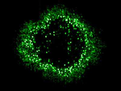
Ring of foveal ganglion cells expressing the calcium indicator GCaMP in vivo.
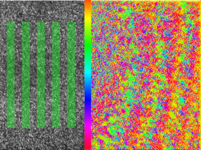
Phase response of retinal ganglion cells to a rolling grating stimulus presented at the fovea.
Optogenetic therapy
Even after photoreceptors have degenerated the ganglion cells, the output neurons of the retina, often persist - can we make these cells light sensitive? This is the idea behind optogenetic vision restoration, where 'optogenes' cause light sensitive proteins to be expressed in cells that don't normally have them and so these cells can be used as photoreceptors instead.
Adaptive optics calcium imaging allows us to evaluate and improve these vision restoration strategies in the living eye by recording activity in foveal retinal ganglion cells. Using the light gated channel 'ChrimsonR' we have demonstrated restored responses in foveal ganglion cells that have lost their photoreceptor input and shown that the expected patterns of activity are generated in response to patterned visual stimuli. We are currently investigating the longevity of optogenetic vision restoration in vivo and evaluating the potential impact of retinal remodelling after photoreceptor loss.
Photoreceptor replacement therapy
To restore naturalistic vision with normal retinal processing it would be desirable to restore vision at the level of the photoreceptors themselves. This requires not only delivering photoreceptor precursor cells capable of maturing and forming synaptic connections into the sub-retinal space, but also that patient's bipolar cell synaptic partners and downstream processing are functionally intact despite many years of photoreceptor loss.
In collaboration with David Gamm at the University of Wisconsin we have been tracking the survival and distribution of fluorescently tagged photoreceptor precursor transplants in the living eye.
Understanding the Fovea
Despite its importance, relatively little is known about the physiology of the fovea because its delicate structure makes it challenging to study electrophysiologically. At Rochester we have been using our unique adaptive optics calcium imaging platform to investigate the fovea in vivo.
By performing calcium imaging while also using the AO system to present high resolution, precisely stabilized visual stimuli, we have been able to map the location of hundreds of retinal ganglion cell (RGC) receptive fields at the fovea. The spatial arrangement of the cone photoreceptors is well preserved in the spatial arrangement of RGCs, despite the dramatic developmental changes which generate the specialized foveal structure. This orderly mapping is of relevance to vision restoration therapies that aim to directly stimulate the retinal ganglion cells themselves. We are also beginning to classify the retinal ganglion cell types found in the fovea using coloured stimuli.
I have a longstanding interest in the physiological optics of the fovea. Extended photoreceptor axons 'Henle Fibres' radiate from the foveal cones to their displaced bipolar cells and interact with macular pigment molecules to produce a radially symmetric polarizing filter which mediates the human perception of the polarization state of light. This visual effect is known as 'Haidinger's brushes'. I've previously explored the threshold and rotational dynamics of the phenomenon using psychophysical testing and am interested in the optical interactions which give rise to it. I often use Haidinger's brushes in outreach activities – almost everyone can discern the polarization state of light with the naked eye!
Selected Publications
- McGregor JE, Kunala K, Xu Z, Murphy P, Godat T, Strazzeri JM, Bateman B, Fischer W, Puthussery T, Williams DR, Merigan WH (2021). Optogenetic therapy restores retinal activity in primate for at least a year following photoreceptor ablation. Molecular Therapy, 1525-0016, https://doi.org/10.1016/j.ymthe.2021.09.014. PDF
- Aboualizadeh E, Phillips MJ, McGregor JE, DiLoreto DA Jr, Strazzeri JM, Dhakal KR, Bateman B, Jager LD, Nilles KL, Stuedemann SA, Ludwig AL, Hunter JJ, Merigan WH, Gamm DM, Williams DR (2020). Imaging Transplanted Photoreceptors in Living Nonhuman Primates with Single-Cell Resolution. Stem Cell Reports. S2213-6711(20)30240-X. doi: 10.1016/j.stemcr.2020.06.019. PDF
- Dhakal KR, Walters S, McGregor JE, Schwarz C, Strazzeri JM, Aboualizadeh E, Bateman B, Huxlin KR, Hunter JJ, Williams DR, Merigan WH (2020). Localized Photoreceptor Ablation Using Femtosecond Pulses Focused With Adaptive Optics. Translational Vision Science & Technology(9), 16. PDF
- McGregor JE, Godat T, Dhakal KR, Parkins K, Strazzeri JM, Bateman BA, Fischer WS, Williams DR, Merigan WH (2020). Optogenetic restoration of retinal ganglion cell activity in the living primate. Nature Communications 11(1), 1703. PDF
- McGregor, J.E. (2019). Restoring vision at the fovea. Current Opinion in Behavioral Sciences 30, 210-216. PDF
- McGregor JE, Yin L, Yang Q, Godat T, Huynh KT, Zhang J, Williams DR, Merigan WH (2018). Functional architecture of the foveola revealed in the living primate. PLoS One 13(11):e0207102. PDF
This material is presented to ensure timely dissemination of scholarly and technical work. Copyright and all rights therein are retained by authors or by other copyright holders. All persons copying this information are expected to adhere to the terms and constraints invoked by each author's copyright. In most cases, these works may not be reposted without the explicit permission of the copyright holder.