Human Retinal Diseases
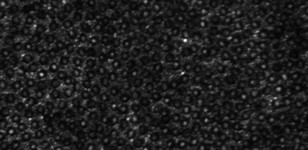
Image of rods and cones
Adaptive optics imaging is a state of the art technology especially suited for non-invasive imaging of the human eye. Individual cells in multiple layers of the retina can be imaged through the dilated pupil. This technology can be used to discover the earliest indicators of retinal disease, follow disease progression over time and to provide quicker endpoints when studying the efficacy of treatments in clinical trials.
Some of the diseases we are studying with adaptive optics imaging are:
AMD Geographic Atrophy
Age-related macular degeneration
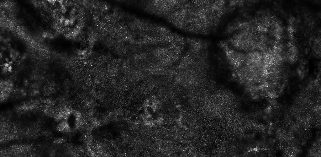
We are studying early disease changes in the photoreceptor and RPE layers and tracking disease progression at a microscopic level. AOSLO has demonstrated intact photoreceptors within geographic atrophy lesions, and has identified changes in the RPE layer within retinal areas which appear normal clinically. By mapping the sequence of disease events, we expect to identify the primary cell layer affected by AMD.
Inherited retinal degenerations
Major discoveries in genetics over the last decade have led to the identification of causative genes in many retinal degenerations. However, the sequence of disease progression from gene defect to cellular abnormality remains poorly understood. AOSLO has demonstrated phenotypic diversity in genetically identical cases of cone rod dystrophy, and has helped to localize the affected cellular layer in retinal diseases, such as fundus albipunctatus, retinitis pigmentosa and Stargardt disease.
Macular telangiectasia
In collaboration with an international study group we are studying this rare condition using AOSLO to identify the natural history of disease progression. We are also participating in the first clinical trial to study a potential therapy for this condition, and developing AOSLO as a potential outcome measure for clinical trials.