In Vivo Retinal Phototoxicity Study
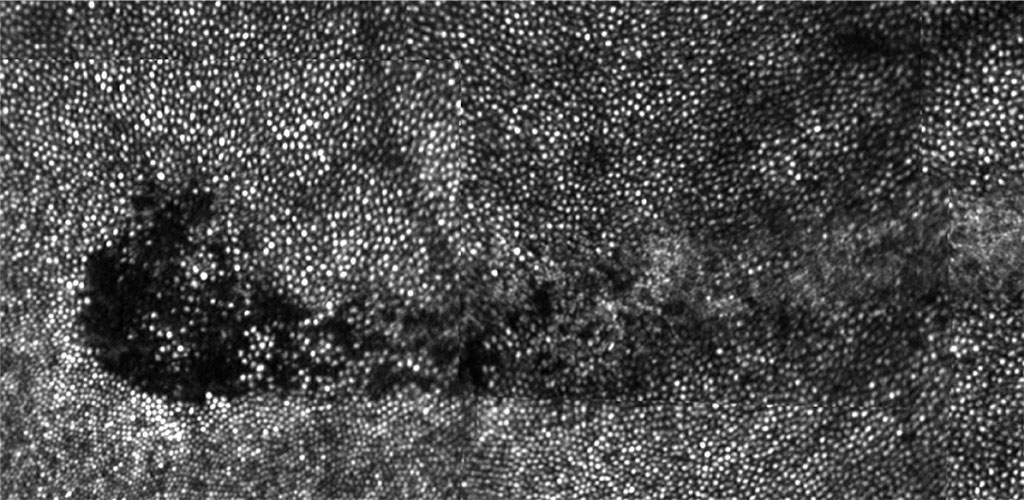
Accidental exposure to a pulsed infrared laser resulted in a patchy loss of photoreceptors colocalized with RPE disruption. The aversion response led to a trail of photoreceptor loss and increased scatter.
Establishing safety limits for light exposure in ophthalmic instruments
We have discovered two new and unexpected effects on the primate retina caused by prolonged exposure to bright visible light (Morgan et al, 2008). The observed effects are autofluorescence (AF) photobleaching and, at higher light levels, retinal pigment epithelium (RPE) disruption (Fig. 1). These effects raise interesting questions about how light interacts with the retina and if they are ultimately deleterious for the eye (Hunter et al, 2012).
Autofluorescence (AF) Photobleaching
Exposures to 568 nm light at irradiances > 5 J/cm2 produce an immediate reduction in lipofuscin AF. This is a transient effect that recovers within several hours. These light levels are up to 2 orders of magnitude below the photochemical maximum exposure (MPE) specified by the ANSI standard for the safe use of lasers (ANSI Z136.1, 2007).
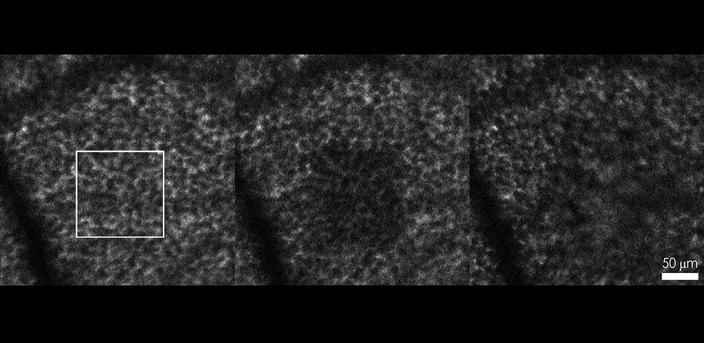
In vivo light damage study: RPE fluorescence pre-exposure, immediately post-exposure, and 6 days post-exposure.
RPE Disruption/Photodamage
Higher exposures (irradiances > 247 J/cm2) to 568 nm light the AF photobleaching effect is followed by a disruption of RPE cell mosaic, seen in RPE fluorescence images in vivo. This RPE disruption occurs at light levels at or slightly below the MPE, which is alarming because the MPE is typically about 10 times below the damage threshold (ANSI Z136.1, 2007). The delay in the appearance of this RPE disruption or photodamage implies that the mechanism of damage is photochemical and not thermal in origin.
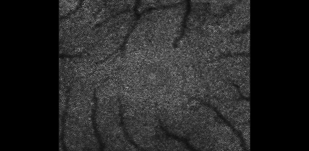
Monkey RPE Mosaic
Publications
- Hunter, J.J., Morgan, J.I.W., Merigan, W.H., Sliney, D.H., Sparrow, J.R., Williams, D.R. (2012). The susceptibility of the retina to photochemical damage from visible light. Progress in Retinal and Eye Research 31(1):28-42. PDF
- Morgan, J.I.W., Hunter, J.J., Williams, D.R., Merigan, W.H. (2009). The reduction of retinal autofluorescence caused by light exposure. Invest Ophthalmol Vis Sci., 50(12):6015-22. PDF
- Morgan, J.I.W., Dubra, A., Wolfe, R., Merigan, W.H., Williams, D.R. (2009). In vivo autofluorescence imaging of the human and macaque retinal pigment epithelial cell mosaic. IOVS, 50(3), 1350-1359. PDF
- Morgan, J.I.W., Hunter, J.J., Masella, B., Wolfe, R., Gray, D.C., Merigan, W.H., Delori, F.C., Williams, D.R. (2008). Light-Induced Retinal Changes Observed with High-Resolution Autofluorescence Imaging of the Retinal Pigment Epithelium. IOVS, 49(8), 3715-3729. PDF