Two-photon Imaging of the Retina
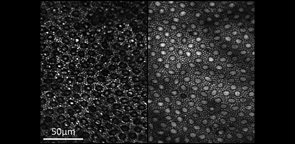
Reflectance (left) and two-photon excited fluorescence (right) image of the photoreceptor mosaic in the living macaque eye. The main source of fluorescence is most likely all-trans-retinol.
Using infrared wavelengths to excite fluorophores with minimum visual stimulation for functional imaging
Two-photon fluorescence imaging has a number of advantages for retinal imaging. Our group was the first to demonstrate two-photon imaging in the living primate eye (Hunter et al, 2011) we are using those results as a stepping stone for the following projects:
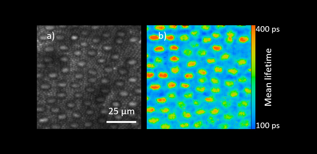
In vivo macaque photoreceptor TPEF intensity (a) and corresponding AOFLIO (b) images displaying mean lifetime
Two-photon Imaging of Endogenous Fluorophores
Two-photon imaging permits the study of molecular species like NADH, FAD, retinol and others. These molecules are direct indicators of cellular function and their excitation regime is in the ultraviolet, which lies outside the transmittance spectrum of the ocular media. Two-photon absorption using infrared light is the only way to excite fluorescence from these molecules and we use this method to study cellular metabolism and the visual cycle in the living eye.
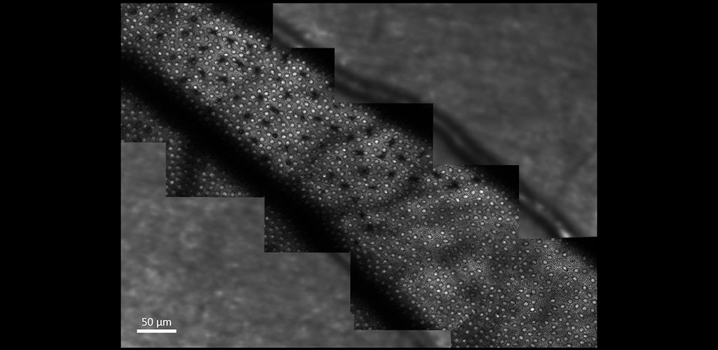
The image shows two-photon excited fluorescence captured from photoreceptors in the living macaque eye where selective S cone damage was induced at different time points.
Adaptive Optics Fluorescence Lifetime Imaging Ophthalmoscopy (AOFLIO)
By combining adaptive optics with fluorescence lifetime imaging, we are able to resolve the fluorescence decay with high resolution both spatially and temporally. Each fluorophore in the retina has a unique fluorescence lifetime which can be modified by environmental factors including enzyme binding. As a result, AOFLIO may be able to help us identify the contribution from retinal fluorophores and how the retinal environment changes when the retina becomes unhealthy. We have shown that cones and rods have different fluorescence lifetimes in the living macaque retina.
Funding Sources
Publications
- Walters S, Schwarz C, Sharma R, Rossi E, Fischer W, DiLoreto D, Strazzeri J, Nelidova D, Roska B, Hunter J, Williams D, and Merigan W (2019). Cellular-scale evaluation of induced photoreceptor degeneration in the living primate eye. Biomed. Opt. Express 10, 66-82. PDF
- Feeks JA & Hunter JJ (2017). Adaptive optics two-photon excited fluorescence lifetime imaging ophthalmoscopy of exogenous fluorophores in mice. Biomed. Opt. Express 8, 2483-2495. PDF
- Sharma R, Yin L, Geng Y, Merigan W, Palczewska G, Palczewski K, Williams D, Hunter J (2013), In vivo two-photon imaging of the mouse retina. Biomed. Opt. Express 4, 1285-1293. PDF
- Hunter, J.J., Masella, B., Dubra, A., Sharma, R., Yin, L., Merigan, W.H., Palczewska, G., Palczewski, K., Williams, D.R. (2011). Images of photoreceptors in living primate eyes using adaptive optics Two-photon ophthalmoscopy. Biomedical Optics Express. 2(1):139-148. PDF