
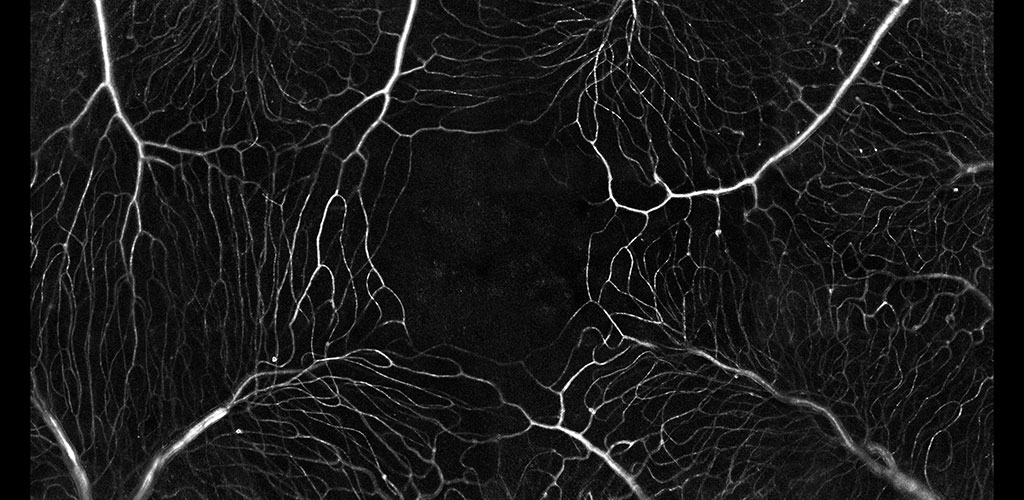
Blood Flow: Vessels of the primate retina color coded by vessel angle.
Blood Flow: The microvascular network imaged without contrast agents. The movement of single blood cells creates a spatio-temporal flicker. Motion contrast imaging reveals active perfusion in the vessels surrounding the fovea.
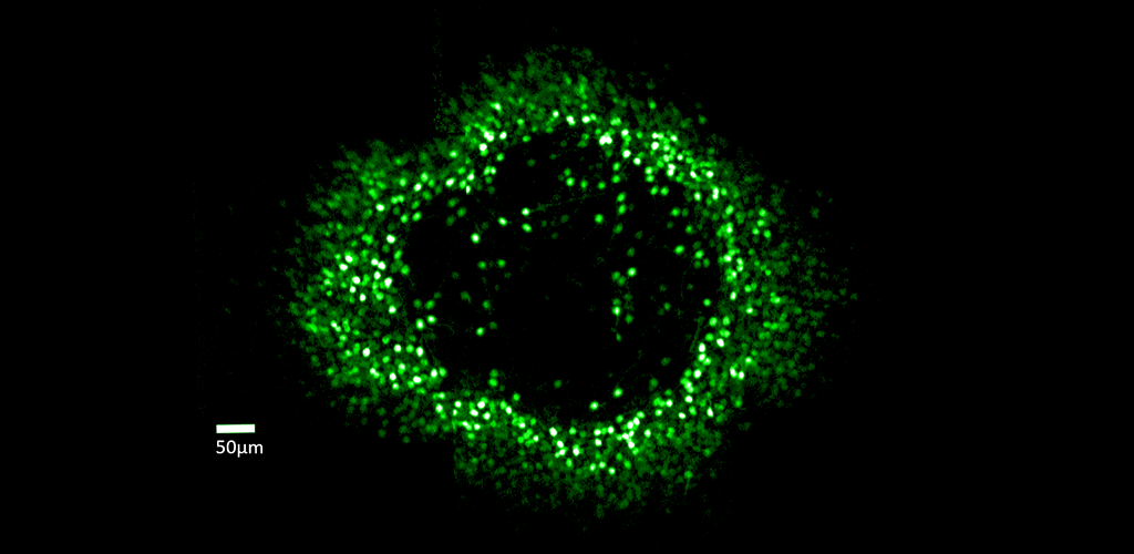
Ganglion Cell Structure & Function: Fluorescent ring of ganglion cells serving the foveal cones. These cells are expressing the calcium indicator GCaMP, which allows cell responses to be monitored optically.
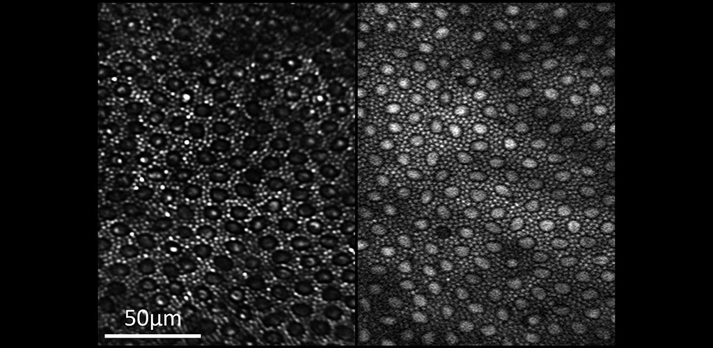
Two-photon Imaging of the Retina: Reflectance (left) and two-photon excited fluorescence (right) image of the photoreceptor mosaic in the living macaque eye. The main source of fluorescence is most likely all-trans-retinol.
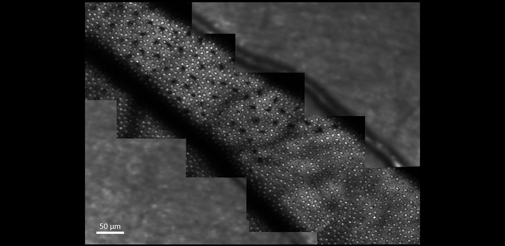
Two-photon Imaging of the Retina: The image shows two-photon excited fluorescence captured from photoreceptors in the living macaque eye where selective S cone damage was induced at different time points.
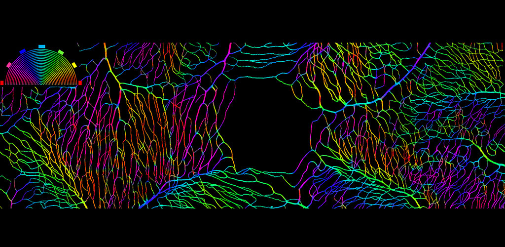
Blood Flow: A binary mask showing perfused vessels surrounding the fovea of the primate. Vessel angle is coded by color.
- Shang F, Dholakia K, Schallek J (2026). One year of hyperglycemia in the Ins2Akita mouse does not impart changes in retinal vascular patterning. PLOS ONE 21(5): e0348363. https://doi.org/10.1371/journal.pone.0348363
- Cai Y, Druszkiewicz E, Patterson SS, Parkins K, McGregor JE, Merigan WH, Fienup JR, Williams DR. Phase diversity improves retinal image quality in adaptive optics scanning light ophthalmoscopy. Biomed. Opt. Express 17, 1767-1781 (2026). https://doi.org/10.1364/BOE.587075
- Murphy PJ, McGregor JE, Xu Z, Yang Q, Merigan W, Williams DR. Optogenetic stimulation of single ganglion cells in the living primate fovea. Elife. 2025 Oct 3;12:RP90050. doi: 10.7554/eLife.90050. PMID: 41041715.