
Blood Flow: Vessels of the primate retina color coded by vessel angle.
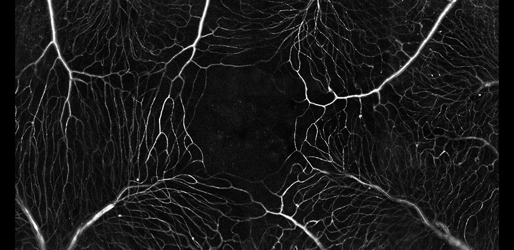
Blood Flow: The microvascular network imaged without contrast agents. The movement of single blood cells creates a spatio-temporal flicker. Motion contrast imaging reveals active perfusion in the vessels surrounding the fovea.
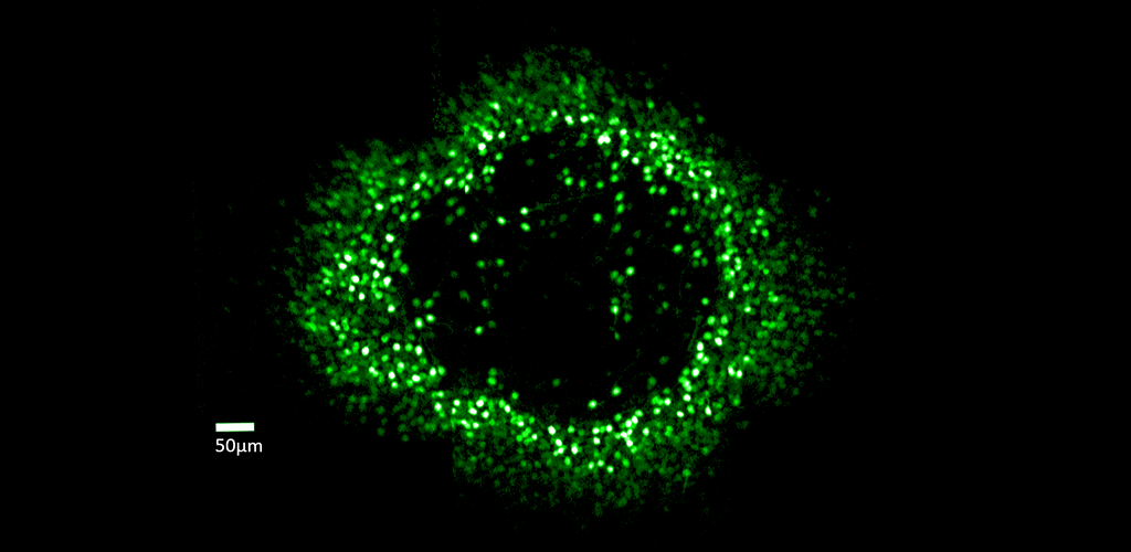
Ganglion Cell Structure & Function: Fluorescent ring of ganglion cells serving the foveal cones. These cells are expressing the calcium indicator GCaMP, which allows cell responses to be monitored optically.
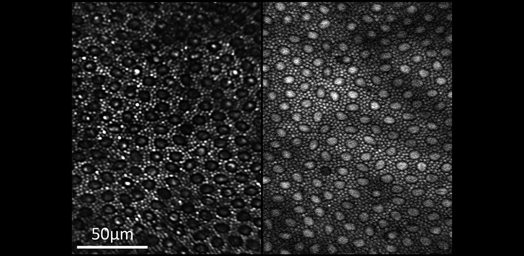
Two-photon Imaging of the Retina: Reflectance (left) and two-photon excited fluorescence (right) image of the photoreceptor mosaic in the living macaque eye. The main source of fluorescence is most likely all-trans-retinol.
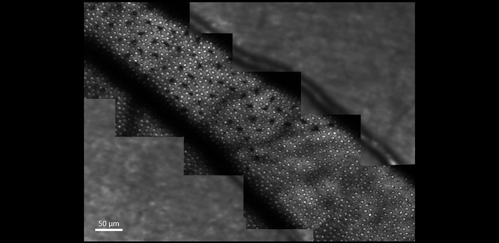
Two-photon Imaging of the Retina: The image shows two-photon excited fluorescence captured from photoreceptors in the living macaque eye where selective S cone damage was induced at different time points.
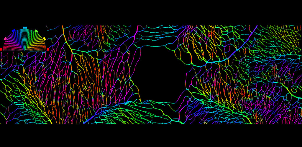
Blood Flow: A binary mask showing perfused vessels surrounding the fovea of the primate. Vessel angle is coded by color.
Overview
ARIA uses adaptive optics imaging of retinal elements to elucidate the structure and function of the human eye. We are a multidisciplinary lab comprised of researchers from the Flaum Eye Institute, the Center for Visual Science and The Institute of Optics at the University of Rochester. This bridge between the engineering and medical worlds, due to the geographic proximity of the world-renowned Optics Institute and the growing Flaum Eye Institute, provides a unique environment for research. Our group is passionate about understanding the physiology underlying healthy and diseased visual systems.
From our research, we have been able to non-invasively see structures such as capillary blood flow and photoreceptor cells in extraordinary detail. This cellular and sub-cellular information is useful in understanding disease progression and prevention, and for informing therapeutic strategies. As methods and systems are advanced, it may be possible to more accurately diagnose retinal diseases at an earlier time point and to more effectively target treatment, as well as to better understand the cellular etymology and pathways of many disorders.