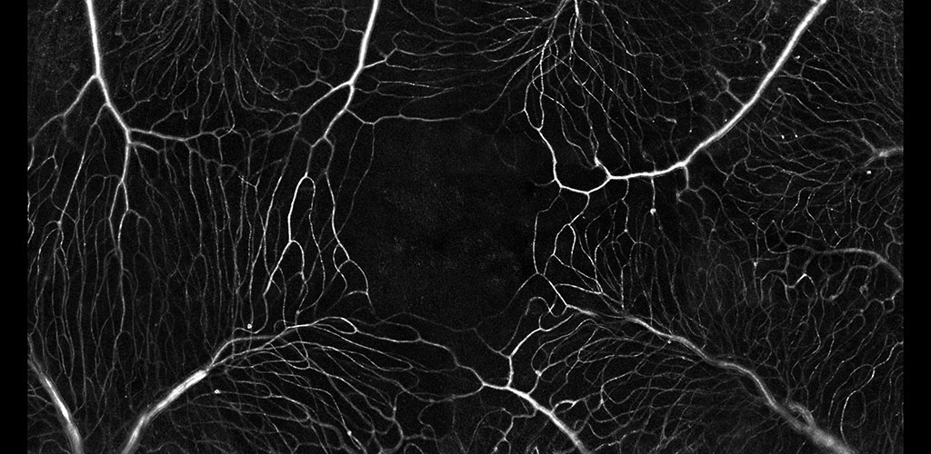
color-coded orientation map of retinal blood vessels in a fundus picture. Each color shows a unique orientation/angle of a vessel

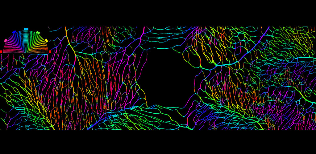
Blood Flow: Vessels of the primate retina color coded by vessel angle.

photo of capillaries of the retina

Blood Flow: The microvascular network imaged without contrast agents. The movement of single blood cells creates a spatio-temporal flicker. Motion contrast imaging reveals active perfusion in the vessels surrounding the fovea.

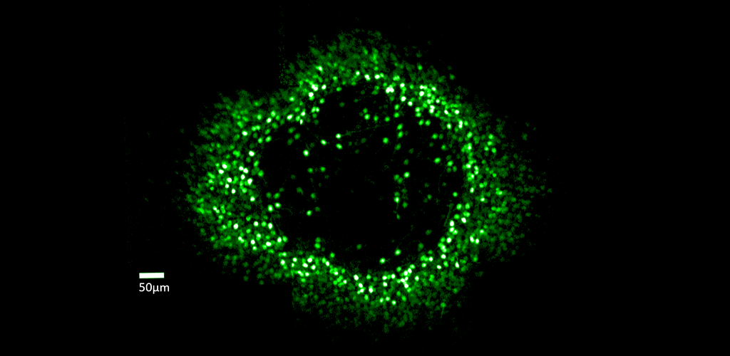
Green ring of cells

Ganglion Cell Structure & Function: Fluorescent ring of ganglion cells serving the foveal cones. These cells are expressing the calcium indicator GCaMP, which allows cell responses to be monitored optically.

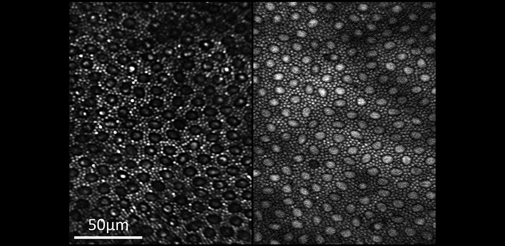
photoreceptor mosaic

Two-photon Imaging of the Retina: Reflectance (left) and two-photon excited fluorescence (right) image of the photoreceptor mosaic in the living macaque eye. The main source of fluorescence is most likely all-trans-retinol.

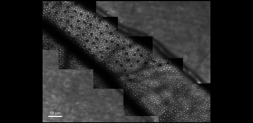
loc88 montage

Two-photon Imaging of the Retina: The image shows two-photon excited fluorescence captured from photoreceptors in the living macaque eye where selective S cone damage was induced at different time points.

Color photo of capillaries of the retina

Blood Flow: A binary mask showing perfused vessels surrounding the fovea of the primate. Vessel angle is coded by color.

Play
Pause

Recent Publications

  • Shang F, Schallek J (2024). Characterization of the Retinal Circulation of the Mouse. Invest Ophthalmol Vis Sci. 2024 Dec 2;65(14):3. doi: 10.1167/iovs.65.14.3. PMID: 39620830.
  • Ashbery D, Baez HC, Kanarr RE, Kunala K, Power D, Chu CJ, Schallek J, McGregor JE; In Vivo Visualization of Intravascular Patrolling Immune Cells in the Primate Eye. Invest. Ophthalmol. Vis. Sci. 2024;65(11):23. https://doi.org/10.1167/iovs.65.11.23.
  • Kunala K, Tang JAH, Parkins K, Hunter JJ (2024). Multispectral label-free in vivo cellular imaging of human retinal pigment epithelium using adaptive optics fluorescence lifetime ophthalmoscopy improves feasibility for low emission analysis and increases sensitivity for detecting changes with age and eccentricity. J. Biomed. Opt. 29(S2) S22707. https://doi.org/10.1117/1.JBO.29.S2.S22707

more publications...